MONDAY, 3 OCTOBER 2011
The discovery and isolation of stem cells has been one of the most exciting and controversial discoveries for the medical sciences in recent decades. Studying these amazing cells, which have the ability to become many different cell types in our body, could significantly improve our ability to repair and replace lost or damaged body parts with fully functional, healthy organs.Much of the interest and conflict surrounding stem cell research relates to embryonic stem cells. These are some of the earliest cells to arise in a developing foetus and are capable of becoming any cell in the body. This gives them huge potential, as it would be possible to take a small group and, with the right signals, generate any part of the body. However, there are many other types of stem cells which are of great interest to biomedical research, but which are generally less highly publicised. These less well-known cells usually produce only a few cell types. Some of them exist even in fully grown adults, whereas embryonic stem cells quickly run out as they change into other cell types.
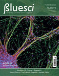
Human neuroepithelial stem cells, which produce some of the earliest cells in the human brain, are just one example and the subject of the cover for this issue. In the brain, neuroepithelial stem cells undergo differentiation, changing from stem cells to neurons (functioning brain cells). The image on the cover shows this process occurring to a small number of cells that were collected and then allowed to grow and divide on a coated plastic surface. This indicates that the process of differentiation is efficient even in a lab environment and so could be used to generate large numbers of brain cells for study or clinical applications. The advantage of starting with neuroepithelial stem cells is that half of the work is done; it would take far more effort to first convert embryonic stem cells to neuroepithelial stem cells and then to use those to generate neurons. Hence, this approach is much faster and more efficient.
The cells in the image have been treated with several coloured markers to allow different cells to be identified. Blue marks the nucleus of all cells, irrespective of cell type. Green cells are those that have differentiated to become neurons. The red colour identifies excitatory neurons: brain cells that act by causing others to increase their signalling activity. Both green and red are visible in the image, indicating that the neuroepithelial stem cells are able to produce several different types of brain cell.
This image was selected for BlueSci by a panel of judges as the winning entry in the Graduate School of Life Sciences Image Competition 2011. It was submitted by Jignesh Tailor, a PhD student working with Dr Austin Smith. Jignesh is keen to develop his work with neuroepithelial stem cells into a means of treating neurodegenerative disorders such as Parkinson’s disease.
The Graduate School of Life Sciences competition includes categories for both images and posters, allowing entrants to discuss their work with many pre-eminent members of the University and to practice their skills at explaining their research to visitors from a wide range of backgrounds. This year, competition was particularly heated and all of the entries were of a very high standard, with many enthusiastic and engaging people getting involved.
The winning entries from all aspects of the competition can be found on the Graduate School of Life Sciences website and other entrants from the image competition will appear throughout this term on our website, www.bluesci.co.uk.
Jonathan Lawson is a first year PhD student at the Gurdon Institute


